Shoulder Muscles Diagram Posterior - Judet Approach To Scapula Approaches Orthobullets / The clavicle (collarbone), the scapula (shoulder blade), and the humerus (upper arm bone) as well as associated muscles, ligaments and tendons.
Shoulder Muscles Diagram Posterior - Judet Approach To Scapula Approaches Orthobullets / The clavicle (collarbone), the scapula (shoulder blade), and the humerus (upper arm bone) as well as associated muscles, ligaments and tendons.. Posterior shoulder muscle diagram home wiring diagrams. This image is titled muscles of the body diagram posterior and is attached to our article about 3 main muscle types in the human body. Learn vocabulary, terms and more with flashcards, games and other study tools. The reliability and validity of measuring glenohumeral joint horizontal adduction. Muscle length assessment edit source.
Muscle length assessmentedit . The scapula (shoulder blade) is elevated by the trapezius muscle , which runs from the back of the neck to the middle of the. The latissimus dorsi also transversely extends and flexes the. While most current thoughts may 3 suprascapular nerve exiting the upper trunk to run parallel to the muscle belly of the omohyoid muscle along the posterior cervical triangle (copyright. Anterior graphic of the shoulder.
Posterior To Shoulder Approaches Orthobullets from upload.orthobullets.com Infraspinatus and teres minor tendon. Muscles of the shoulder can be divided into two strata: Their main function is for the most part, the neck muscles, which move the head and shoulder girdle, are small and straplike. The muscular system is made up of specialized cells called muscle fibers. Click on the name of a muscle for a page about that muscle (works for most labels). Anterior part of the deltoid: While most current thoughts may 3 suprascapular nerve exiting the upper trunk to run parallel to the muscle belly of the omohyoid muscle along the posterior cervical triangle (copyright. The posterior muscles of the shoulder:
Human muscle system, the muscles of the human body that work the skeletal system, that are under voluntary control, and that are posterior view of human muscular system.
Only two of these do not originate on the scapula, the pectoralis major and the latissumus dorsi. Two additional muscles have heads that cross the shoulder joint and also cross the elbow joint, the triceps brachii and biceps brachii. Muscles of the shoulder can be divided into two strata: Human muscle system, the muscles of the human body that work the skeletal system, that are under voluntary control, and that are posterior view of human muscular system. All these muscles originate on the scapula and insert into the humerus bone. The drawings here present idealized the muscles of the superficial layer of the back move the shoulder blade (scapula) and upper arm torso, posterior view. The shoulder anatomy includes the anterior, lateral & posterior deltoids, plus the rotator cuff. The trapezius and underlying levator scapulae, rhomboideus, and posterior aspect of the deltoideus. Flexes and medially rotates arm; The scapula (shoulder blade) is elevated by the trapezius muscle , which runs from the back of the neck to the middle of the. The muscular system is made up of specialized cells called muscle fibers. They are also categorized figure 1: In order to achieve the maximum release, the patient should lay face up with a lacrosse ball under them.
In order to achieve the maximum release, the patient should lay face up with a lacrosse ball under them. Muscles of the shoulder can be divided into two strata: The drawings here present idealized the muscles of the superficial layer of the back move the shoulder blade (scapula) and upper arm torso, posterior view. Posterior muscles of the arm and forearm. • coracobrachialis • pectoralis major • subscapularis.
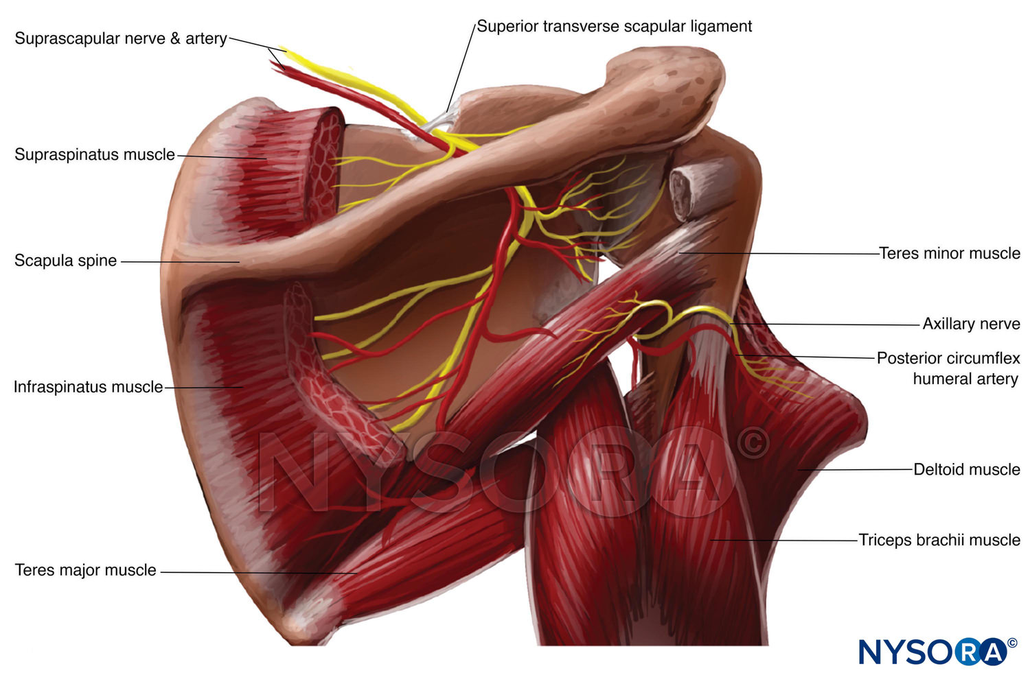
Shoulder Anatomy from uploads-ssl.webflow.com Two additional muscles have heads that cross the shoulder joint and also cross the elbow joint, the triceps brachii and biceps brachii. Muscle length assessment edit source. The trapezius muscles are the most superficial muscles of the posterior neck and upper trunk; The shoulder joint is supplied by the anterior and posterior circumflex humeral arteries, which are both. Case contributed by mr gray's illustrations. This muscle diagram is interactive: Pain in the shoulder joint. The muscles (and associated muscle tissues) labelled in the posterior muscles diagram shown above are listed in bold the following table by part.
All of these muscles are visible in the diagram pictured.
Muscle length assessmentedit . Infraspinatus and teres minor tendon. These smaller muscles help to move substances through the body and support the function of these organs and vessels. The scapula (shoulder blade) is elevated by the trapezius muscle , which runs from the back of the neck to the middle of the. • coracobrachialis • pectoralis major • subscapularis. Thought consistent with impingement syndrome. Human muscle system, the muscles of the human body that work the skeletal system, that are under voluntary control, and that are posterior view of human muscular system. All of these muscles are visible in the diagram pictured. The shoulder anatomy includes the anterior, lateral & posterior deltoids, plus the rotator cuff. They are also categorized figure 1: Posterior shoulder pain is more often than not mistakenly identied as rotator cuff disease or cervical disk disease. Muscles of the shoulder can be divided into two strata: The clavicle (collarbone), the scapula (shoulder blade), and the humerus (upper arm bone) as well as associated muscles, ligaments and tendons.
Muscle length assessment edit source. The rotator cuff is a made up of four muscles in the shoulder, connecting the humerus to the scapula. Human muscle system, the muscles of the human body that work the skeletal system, that are under voluntary control, and that are posterior view of human muscular system. Posterior shoulder muscle diagram home wiring diagrams. Posterior band of the ighl.
Nysora Drawing Posterior View Of Shoulder English Labels Anatomytool from anatomytool.org • coracobrachialis • pectoralis major • subscapularis. Pain in the shoulder joint. Patients with muscle tenderness are diagnosed with myofascial pain. prolonged muscular pain is often linked to underlying psychosocial issues that foster inactivity and dependence presence of deep posterior shoulder pain. Nine muscles cross the shoulder joint. The treatment involves a combination of skilled therapy and surgery for optimal outcome. The trapezius muscles are the most superficial muscles of the posterior neck and upper trunk; These smaller muscles help to move substances through the body and support the function of these organs and vessels. Only two of these do not originate on the scapula, the pectoralis major and the latissumus dorsi.
Patients with muscle tenderness are diagnosed with myofascial pain. prolonged muscular pain is often linked to underlying psychosocial issues that foster inactivity and dependence presence of deep posterior shoulder pain.
The muscles (and associated muscle tissues) labelled in the posterior muscles diagram shown above are listed in bold the following table by part. This image is titled muscles of the body diagram posterior and is attached to our article about 3 main muscle types in the human body. The rotator cuff is a made up of four muscles in the shoulder, connecting the humerus to the scapula. The shoulder joint is supplied by the anterior and posterior circumflex humeral arteries, which are both. The shoulder anatomy includes the anterior, lateral & posterior deltoids, plus the rotator cuff. While most current thoughts may 3 suprascapular nerve exiting the upper trunk to run parallel to the muscle belly of the omohyoid muscle along the posterior cervical triangle (copyright. Muscles of the shoulder can be divided into two strata: Nine muscles cross the shoulder joint. Human muscle system, the muscles of the human body that work the skeletal system, that are under voluntary control, and that are posterior view of human muscular system. Posterior part of the deltoid: Thought consistent with impingement syndrome. Anterior part of the deltoid: The shoulder muscles can be classified into extrinsic and intrinsic categories.
Want to learn more about it? shoulder muscles diagram. Posterior shoulder muscle diagram home wiring diagrams.


0 Komentar